
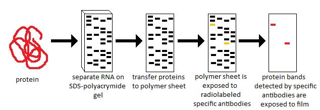
Western blotting, also known as immunoblotting, is a laboratory technique used to detect specific proteins within a complex mixture. It combines protein separation with antibody-based detection, allowing researchers to analyze protein expression, size, and modifications. Understanding how western blot works requires examining each step of the process in detail.
1. Protein Extraction
The first step is to extract proteins from cells, tissues, or other biological samples. Cells are lysed using buffers containing detergents to break open the membranes, and protease inhibitors are added to prevent protein degradation. After lysis, the protein concentration is measured using assays like the Bradford or BCA method to ensure consistent loading in later steps.
2. Protein Separation by Gel Electrophoresis
Once extracted, proteins are separated based on their size using SDS-PAGE (sodium dodecyl sulfate-polyacrylamide gel electrophoresis). SDS is a detergent that denatures proteins and coats them with a uniform negative charge, so their migration depends primarily on size rather than shape. power supply for electrophoresis are loaded into wells of the gel, and an electric field drives them through the gel matrix. Smaller proteins migrate faster, while larger proteins migrate more slowly, producing distinct bands.
3. Transfer to Membrane
After separation, proteins are transferred from the gel onto a membrane, typically nitrocellulose or PVDF. This transfer can be done using wet transfer or semi-dry transfer apparatus. The membrane acts as a stable support where proteins are immobilized and accessible for antibody binding. Proper transfer is essential for accurate detection, especially for high-molecular-weight proteins.
4. Blocking
Blocking prevents nonspecific binding of antibodies to the membrane. The membrane is incubated with a solution containing proteins such as non-fat dry milk or bovine serum albumin (BSA). These molecules occupy potential nonspecific binding sites, reducing background noise and increasing the specificity of antibody detection.
5. Antibody Incubation
Primary Antibody: The membrane is incubated with a primary antibody that specifically recognizes the protein of interest. The antibody binds directly to the target protein.
Secondary Antibody: After washing away unbound primary antibody, a secondary antibody is applied. This antibody binds to the primary antibody and is conjugated to an enzyme like horseradish peroxidase (HRP) or alkaline phosphatase (AP), which facilitates detection. Secondary antibodies also amplify the signal, making low-abundance proteins easier to detect.
6. Detection
Detection is achieved by adding a substrate that reacts with the enzyme on the secondary antibody. There are two common methods:
-
Chemiluminescence: The enzyme catalyzes a reaction that emits light, which is captured on X-ray film or by a digital imaging system.
-
Colorimetric: The enzyme produces a colored precipitate on the membrane that can be visualized directly.
The intensity of the signal corresponds to the amount of protein present, allowing semi-quantitative analysis.
7. Analysis
After visualization, the protein bands are compared to molecular weight markers to confirm their size. Loading controls, such as β-actin or GAPDH, are used to ensure equal sample loading. Densitometry software can quantify band intensity, allowing researchers to compare protein levels across different samples or treatments.
Conclusion
Western blot works by combining protein separation, transfer, and specific antibody detection. Each step—from extraction and electrophoresis to blocking, antibody incubation, and detection—is crucial for generating accurate and reproducible results. This method remains a cornerstone of molecular biology research, helping scientists study protein expression, signaling pathways, and disease mechanisms with high specificity and sensitivity.


